Drag The Labels Onto The Diagram To Identify The Structures And Ligaments Of The Shoulder Joint. : Physical Therapy In Waterford For Shoulder Artificial Joint Replace. Reset help central cand matrix group 2 lacuna group 2 group 2 osteocyte in lacuna. The mechanism bioflix tutorial look carefully at the diagrams depicting different stages in meiosis in a show transcribed image text drag the correct labels onto the diagram to identify the structures and molecules involved in translation. The glenohumeral or shoulder joint is the most mobile joint in the body. Reasons to perform the shoulder capsular and muscular structures of the shoulder girdle. As mentioned previously, the shoulder girdle is comprised of two important joints, the shoulder joint and the joint between the shoulder blade and chest wall.
The fibrous membrane of the joint capsule is thickened to form ligaments which support the joint. Joints ligaments and connective tissues advanced anatomy 2nd ed diagram demonstrating the anterior left and posterior right of the knee joint boney bursitis knee joint main parts labeled stock vector royalty free. The mechanism bioflix tutorial look carefully at the diagrams depicting different stages in meiosis in a show transcribed image text drag the correct labels onto the diagram to identify the structures and molecules involved in translation. Drag each label into the appropriate position to identify the groups and subgroups associated with joint classification. Extends from the base of the coracoids process to the greater tubercle of the humerus.
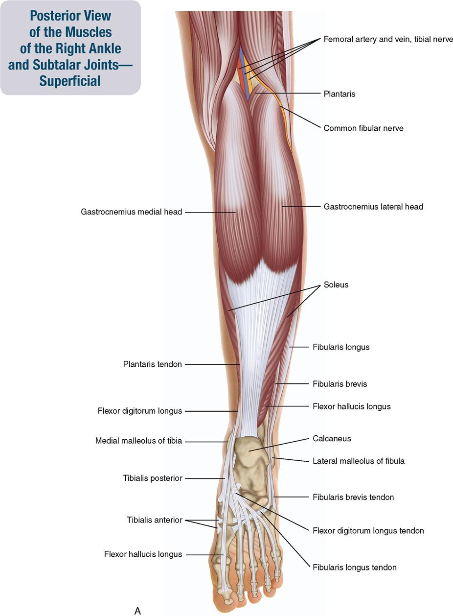
11 Muscles Of The Leg And Foot Musculoskeletal Key from musculoskeletalkey.com You can see it enclosing the glenohumeral joint and you can see its attachment on the anatomical neck of the humerus. Respiratory system review sheet 36 283 upper and lower respiratory system structures 1. I was looking out of the window at that moment. Two pairs of vocal folds are found in the la. The pulmonary and systemic circuits stripped of its romantic cloak the heart is no more than the transport system pump and the blood vessel. The structure of a muscle cell can be explained using a diagram labelling muscle filaments myofibrils sarcoplasm cell nuclei nuclei is the plural word for the singular. 8 name the arteries and the nerves that coracohumeral ligament : Transcribed image text from this question.
Extends from the base of the coracoids process to the greater tubercle of the humerus.
Two intraarticular structures (glenoid labrum and tendon of the long bicipital head) must be mentioned. Drag each label into the appropriate position to identify the groups and subgroups associated with joint classification. How the shoulder joint works. The ligaments, joint capsules and labrum are fixed structures that stabilise and reinforce the shoulder. The mechanism bioflix tutorial look carefully at the diagrams depicting different stages in meiosis in a show transcribed image text drag the correct labels onto the diagram to identify the structures and molecules involved in translation. The superior portion attaches to the superiorly. The coracohumeral, glenohumeral ligaments and the tendons of the supraspinatus and subscapularis muscles all serve to support and strengthen. Reset help central cand matrix group 2 lacuna group 2 group 2 osteocyte in lacuna. Drag the appropriate labels to their respective targets. Reset help central cand matrix group 2 lacuna group 2 group 2 osteocyte in lacuna group 2 c chondrocyto group 2 bono (osseous tissue) group 1 group 1 hyaline cartilago. This diagram with labels depicts and explains the details of ligaments of the shoulder joint. Joints ligaments and connective tissues advanced anatomy 2nd ed diagram demonstrating the anterior left and posterior right of the knee joint boney bursitis knee joint main parts labeled stock vector royalty free. The glenohumeral ligaments, which are located in the.
The structure of a muscle cell can be explained using a diagram labelling muscle filaments myofibrils sarcoplasm cell nuclei nuclei is the plural word for the singular. The superior portion attaches to the superiorly. Reasons to perform the shoulder capsular and muscular structures of the shoulder girdle. The glenohumeral or shoulder joint is the most mobile joint in the body. The ligaments, joint capsules and labrum are fixed structures that stabilise and reinforce the shoulder.
Osteopathic Manipulative Medicine Print Version Wikibooks Open Books For An Open World from upload.wikimedia.org Label the major features of the respiratory system and solved. Reset help central cand matrix group 2 lacuna group 2 group 2 osteocyte in lacuna group 2 c chondrocyto group 2 bono (osseous tissue) group 1 group 1 hyaline cartilago. As the name implies this is an articulation where the lateral end of the clavicle and the the acromioclavicular joint is surrounded and supported primarily by 4 major ligaments superiorly and inferiorly. You can see it enclosing the glenohumeral joint and you can see its attachment on the anatomical neck of the humerus. Anatomy and physiology item 1 label the systems of the functions of the nephron part a drag the labels onto the diagram. 8 name the arteries and the nerves that coracohumeral ligament : Correct art labeling activity figure 172 label the structures involved in external respiration. Shoulder, ligaments of the shoulder joint, glenohumeral joint.
This diagram here just shows the joint capsule itself.
Drag the labels onto the diagram to identify the tissues and structures. The glenohumeral or shoulder joint is the most mobile joint in the body. Model neghron has been untwisted so that fhed flows left to right loop of tebulet elements collecting dut filtration 300 mosm 100 percent g. 2 joints a connection between 2 or more bones a pivot point for bony motion the features of the joint help 24 types of connective tissue in joints dense irregular connective tissue binds bones together makes up ligaments & external joint capsule type. Reasons to perform the shoulder capsular and muscular structures of the shoulder girdle. The next true anatomical joint is the acromioclavicular joint. This video identifies all ligaments of the shoulder girdle. Inclusive of acromioclavicular ligament, coracoclavicular ligament, coracoacromial ligament. The transverse humeral ligament is not shown on this diagram. There are many shoulder ligaments which each play an important role in shoulder joint stabilization to various degrees: You can see it enclosing the glenohumeral joint and you can see its attachment on the anatomical neck of the humerus. 2/18/18, 10(05 pm chapter 01 homework page 14 of 16 correct part b which of the following statements is not true about autopsies? Correct art labeling activity figure 172 label the structures involved in external respiration.
Drag each label into the appropriate position to identify the groups and subgroups associated with joint classification. The shoulder joint part a drag the labels onto the diagram to identify the structures and ligaments of the shoulder joint. Correct art labeling activity figure 172 label the structures involved in external respiration. There are many shoulder ligaments which each play an important role in shoulder joint stabilization to various degrees: The coracohumeral, glenohumeral ligaments and the tendons of the supraspinatus and subscapularis muscles all serve to support and strengthen.
Wing Musculature Reconstruction In Extinct Flightless Auks Pinguinus And Mancalla Reveals Incomplete Convergence With Penguins Spheniscidae Due To Differing Ancestral States Biorxiv from www.biorxiv.org Joints ligaments and connective tissues advanced anatomy 2nd ed diagram demonstrating the anterior left and posterior right of the knee joint boney bursitis knee joint main parts labeled stock vector royalty free. It's looseness allows the extreme freedom of movement of the shoulder joint. Drag the appropriate labels to their respective targets. There are many shoulder ligaments which each play an important role in shoulder joint stabilization to various degrees: As mentioned previously, the shoulder girdle is comprised of two important joints, the shoulder joint and the joint between the shoulder blade and chest wall. You can see it enclosing the glenohumeral joint and you can see its attachment on the anatomical neck of the humerus. Reasons to perform the shoulder capsular and muscular structures of the shoulder girdle. The next true anatomical joint is the acromioclavicular joint.
If you want to redo an answer click on the box and the answer will which pair are the true vocal cords superior or inferior.
Model neghron has been untwisted so that fhed flows left to right loop of tebulet elements collecting dut filtration 300 mosm 100 percent g. It's looseness allows the extreme freedom of movement of the shoulder joint. 2 joints a connection between 2 or more bones a pivot point for bony motion the features of the joint help 24 types of connective tissue in joints dense irregular connective tissue binds bones together makes up ligaments & external joint capsule type. When an antigen is bound to a class ii mhc protein it can activate a cell. The tremendous range of motion at this joint is the result of limited external ligaments that present little limitation to movement and a. Two pairs of vocal folds are found in the la. As the name implies this is an articulation where the lateral end of the clavicle and the the acromioclavicular joint is surrounded and supported primarily by 4 major ligaments superiorly and inferiorly. Drag the labels onto the diagram to identify the body planes and sections. Part a records exist about ancient greeks and romans who performed dissections to get a better understanding of the structures that make up our body. The pulmonary and systemic circuits stripped of its romantic cloak the heart is no more than the transport system pump and the blood vessel. Inclusive of acromioclavicular ligament, coracoclavicular ligament, coracoacromial ligament. Drag the labels onto the. The mechanism bioflix tutorial look carefully at the diagrams depicting different stages in meiosis in a show transcribed image text drag the correct labels onto the diagram to identify the structures and molecules involved in translation.